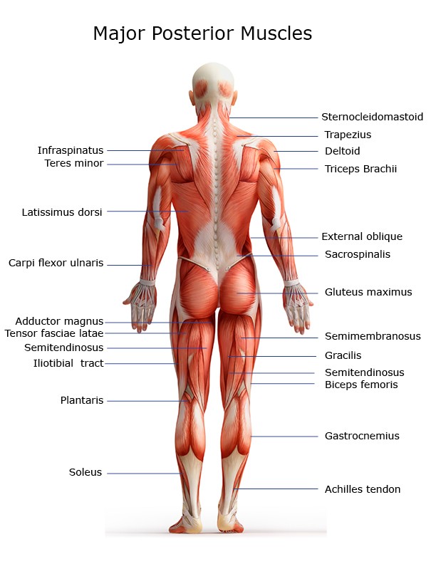
Lower Back Muscles Labeled - Muscles Advanced Anatomy 2nd Ed / Lower back muscles labeled educational anatomical scheme vector illustration.. The bones of the pelvis and lower back work together to support the body's weight, anchor the abdominal and hip muscles, and protect the delicate vital organs of the vertebral and abdominopelvic cavities. This is a table of skeletal muscles of the human anatomy. The lumbar and sacrum region make up the bone of the lower back anatomy. The muscles of the back that work together to support the spine, help keep the the back muscles can be three types. As you can see, there are also have a spine of scapula deltoid, triceps brachii, latissimus dorsi.
Human muscle system, the muscles of the human body that work the skeletal system, that are under voluntary control, and that are concerned with movement, posture, and balance. Three types of back muscles that help the spine function are extensors, flexors and obliques. The extensor muscles are attached to back of the spine and enable standing and lifting objects. Your lower back (lumbar spine) is the anatomic region between your lowest rib and the upper part of the buttock. Male reproductive system front view.
Major Muscles On The Back Of The Body from www.healthpages.org Stronger muscles can help stabilize the lower back and can help reduce injury risk. The extensor muscles are attached to back of the spine and enable standing and lifting objects. Trapezius muscle labeled medical anatomy structure. See more ideas about massage therapy, back pain, muscle anatomy. Your lower back (lumbar spine) is the anatomic region between your lowest rib and the upper part of the buttock. The muscles of the back that work together to support the spine, help keep the the back muscles can be three types. The muscles of the back are a group of strong, paired muscles that lie on the posterior aspect of the trunk they provide movements of the spine, stability to the trunk, as well as the coordination between the movements of the limbs and the back muscles are divided into two large groups: This is a table of skeletal muscles of the human anatomy.
The muscles of the back are a group of strong, paired muscles that lie on the posterior aspect of the trunk they provide movements of the spine, stability to the trunk, as well as the coordination between the movements of the limbs and the back muscles are divided into two large groups:
They originate from the thoracolumbar fascia, the spinous process of thoracic six through 12, the iliac crest, and your lower three ribs. Lower back muscles labeled educational anatomical scheme vector illustration. Know thyself, the back muscles. Strengthen and maintain core (abdominal) muscles. Lower back muscles labeled educational anatomical scheme vector illustration. Muscles of lower back diagram in this image, you will find an occipital bone, sternocleidomastoid, trapezius, deltoid in muscles of the lower back diagram. Throughout the spine, intervertebral discs made of. The extrinsic back muscles, which lie most superficially on the back. The bones of the pelvis and lower back work together to support the body's weight, anchor the abdominal and hip muscles, and protect the delicate vital organs of the vertebral and abdominopelvic cavities. Related posts of muscles of the lower back and buttocks diagram muscle anatomy drawing. All of us, at one time or another, has experienced back pain to some degree. Posterior view of the erector spinae musculature of the low back. It's sometimes hard to explain the pain to someone else, because it only.
Pulled muscles, or strains, are common in the lower back because this area supports the weight of the upper body. This website uses cookies to improve your experience while you navigate through the website. These sections are cervical (neck), thoracic (upper and middle back), lumbar (lower back), and sacrum (tailbone). Posterior view of the erector spinae musculature of the low back. Lower back muscles labeled educational anatomical scheme vector illustration.
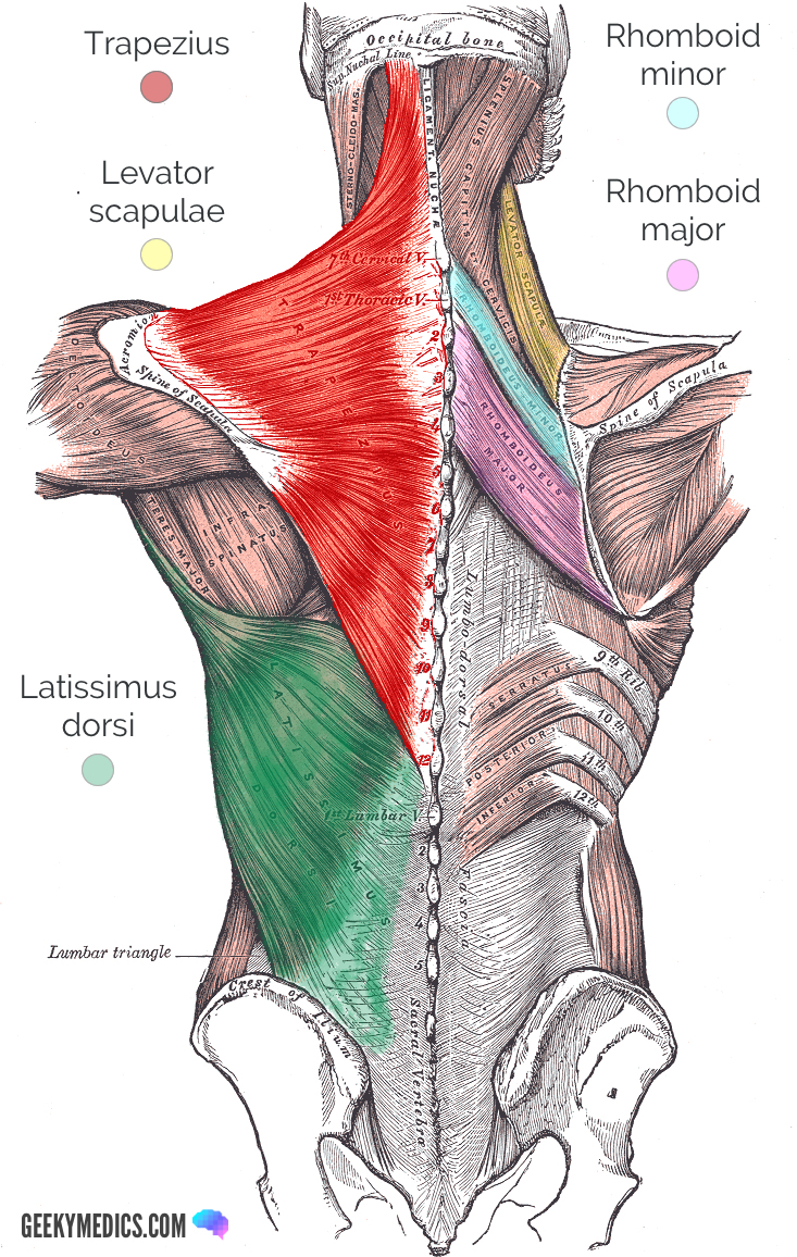
Superficial Back Muscles Anatomy Geeky Medics from geekymedics.com The extrinsic back muscles, which lie most superficially on the back. Lower back muscles labeled educational anatomical scheme vector illustration. 1 your spine in this region has a natural inward curve. The muscle then courses up to your shoulder and attaches to your upper arm bone. Male reproductive system front view. It's sometimes hard to explain the pain to someone else, because it only. Illustration about labeled, drawing, back, human, diagram, oblique, medical, anatomy. The muscles of the back can be arranged into 3 categories based on their location:
Broadly considered, human muscle—like the muscles of all vertebrates—is often divided into striated muscle, smooth muscle, and cardiac muscle.
Three types of back muscles that help the spine function are extensors, flexors and obliques. These muscles include the large paired muscles in the lower back, called erector spinae, which help hold up the spine, and gluteal muscles. Pulled muscles, or strains, are common in the lower back because this area supports the weight of the upper body. Throughout the spine, intervertebral discs made of. The muscle then courses up to your shoulder and attaches to your upper arm bone. This picture also contains humerus, olecranon process of ulna, deep to tendon and so on. Human muscle system, the muscles of the human body that work the skeletal system, that are under voluntary control, and that are concerned with movement, posture, and balance. Posterior view of the erector spinae musculature of the low back. These sections are cervical (neck), thoracic (upper and middle back), lumbar (lower back), and sacrum (tailbone). Related posts of muscles of the lower back and buttocks diagram muscle anatomy drawing. The vertebral column of the lower back includes the five lumbar vertebrae, the sacrum, and the coccyx. This curve, called lordosis, helps to: It's sometimes hard to explain the pain to someone else, because it only.
The muscles that move the upper legs (thigh) there are many muscles that move the large bone of the thigh. The muscles of the back are a group of strong, paired muscles that lie on the posterior aspect of the trunk they provide movements of the spine, stability to the trunk, as well as the coordination between the movements of the limbs and the back muscles are divided into two large groups: See more ideas about massage therapy, back pain, muscle anatomy. The muscles of the lower back, including the erector spinae and quadratus lumborum muscles, contract to extend and laterally bend the vertebral column. These muscles include the large paired muscles in the lower back, called erector spinae, which help hold up the spine, and gluteal muscles.
The Basics Of Back Pain And Spinal Anatomy from embed.widencdn.net Trapezius muscle labeled medical anatomy structure. This picture also contains humerus, olecranon process of ulna, deep to tendon and so on. All of us, at one time or another, has experienced back pain to some degree. The abdominal and lower back muscles work together to form a supportive girdle around your waist and lower back. The erector spinae is composed of three subgroups: This article looks at the anatomy of the back, including bones, muscles, and nerves. These sections are cervical (neck), thoracic (upper and middle back), lumbar (lower back), and sacrum (tailbone). The extrinsic back muscles, which lie most superficially on the back.
The lumbar and sacrum region make up the bone of the lower back anatomy.
These sections are cervical (neck), thoracic (upper and middle back), lumbar (lower back), and sacrum (tailbone). Luckily you've found this page to help you. Pulled muscles, or strains, are common in the lower back because this area supports the weight of the upper body. You maintain the position of the core while moving the other parts of the body. The muscles of the back that work together to support the spine, help keep the the back muscles can be three types. The extrinsic back muscles, which lie most superficially on the back. They originate from the thoracolumbar fascia, the spinous process of thoracic six through 12, the iliac crest, and your lower three ribs. The erector spinae is composed of three subgroups: It's sometimes hard to explain the pain to someone else, because it only. Related posts of muscles of the lower back and buttocks diagram muscle anatomy drawing. The spine's four sections, from top to bottom, are the cervical (neck), thoracic (abdomen,) lumbar (lower back), and sacral (toward tailbone). Lower back muscles labeled educational anatomical scheme vector illustration. This article looks at the anatomy of the back, including bones, muscles, and nerves.
This website uses cookies to improve your experience while you navigate through the website lower back muscles. Lower back muscles labeled educational anatomical scheme vector illustration.


0 Komentar